
Understanding Veterinary Radiography
The Role of Diagnostic Imaging in Animal Medicine
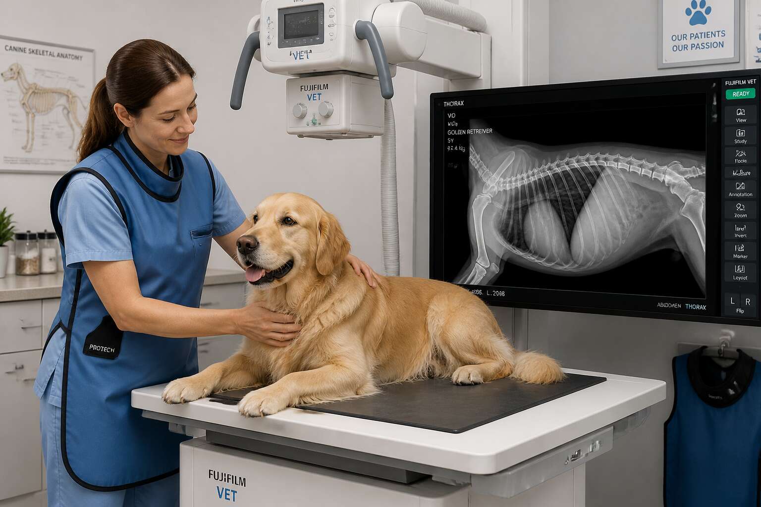
Veterinary radiography has been around for more than a hundred years, yet most pet owners still cannot read a film. That is where the expertise at a vet clinic xray comes in. A single image reveals fractures, fluid accumulation, and foreign bodies with clarity that physical examination cannot match.
Modern digital radiography offers distinct advantages over older film systems. Images appear in seconds, exposure errors drop significantly, and files integrate directly into patient records. Radiographs are commonly requested for suspected:
- Bone fractures or joint damage
- Heart and lung abnormalities
- Bladder or intestinal blockages
The value extends beyond diagnosis. Serial radiographs track healing progress, confirm surgical placements, and guide treatment adjustments over time. Pet owners increasingly expect access to a vet clinic xray when symptoms appear, and South African practices are responding by investing in better equipment. In my experience, owners are often surprised by how quickly the whole process moves.
Common Types of Imaging Equipment Used by Veterinarians
Ever watched a Great Dane try to sit still for a chest X-ray? Patience has its limits. That’s why modern vet clinic xray setups rely on hardware.
Digital radiography captures images in milliseconds. Computed radiography, using cassettes and a reader, works well in mobile units. Ultrasound offers real-time soft tissue views, while CT scans provide 3D detail for complex spinal cases. Each tool has a role, and the best teams mix them. That often includes:
- High-frequency generators for sharper images
- Wireless flat panel detectors for speed
These components make the procedure faster and more precise. The hardest part is convincing your pet to hold still.
How Veterinary Imaging Differs from Human Radiography
A human patient holds still on command. A cat with a grudge holds its breath instead. That single distinction drives the entire philosophy of veterinary radiography.
Human radiography assumes cooperation. The subject adjusts their position, inhales, and stays still. Veterinary imaging requires the radiographer to adapt to a subject that refuses to cooperate. Animals arrive with fur, feathers, or scales, each affecting the beam. Their anatomical proportions differ sharply from ours, so exposure settings are never routine. The vet clinic xray team must compensate for a dog’s thick chest wall or a bird’s delicate ribs.
- Calibration follows the breed, not the birthdate.
- Restraint devices replace verbal instructions.
These adjustments transform an X-ray exam into a custom performance for every species.
Signs Your Pet May Need Diagnostic Imaging
Unexplained Lameness or Mobility Issues
Your dog’s three-legged shuffle is not a new dance craze. Unexplained lameness that lingers beyond a day often means something deeper. A vet clinic xray can reveal fractures, joint disease, or bone tumours that a physical exam cannot feel.
Watch for these red flags:
- Reluctance to climb stairs or jump
- Audible whimpers when rising
- Dragging a limb during walks
If your cat suddenly refuses to use the litter box due to mobility pain, that also counts. Persistent stiffness after rest, rather than after exercise, warrants imaging. Do not assume it is age. Your pet might be hiding discomfort, but the xray will expose the truth.
Persistent Vomiting or Gastrointestinal Distress
Persistent vomiting is more than a mess. It often signals an obstruction, a foreign body, or a twisted organ. A single episode after a questionable snack is normal. Repeated bouts, especially with visible distress, require more than a bland diet.
- Vomiting that resumes hours after a meal
- Blood-streaked stool or dark, tarry waste
- A bloated, taut abdomen that feels painful to touch
- Restlessness and refusal to settle
These signs point to a physical problem, not a behavioral one. A physical exam cannot see through layers of tissue and fluid. That is where a vet clinic xray becomes essential. It can reveal a swallowed object or an intestinal twist before the situation turns critical.
When you cannot keep food down for a day, your body speaks. Listen with imaging.
Potential Foreign Body Ingestion
A dog that swallows a peach pit rarely shows distress immediately. The object may sit in the stomach for days before causing trouble. Subtle changes in behaviour are often the first clue.
Watch for these early signs:
- Pawing at the mouth or excessive lip licking
- Refusing food but drinking water
- Straining to pass stool or producing only small amounts
- Whining when the abdomen is touched
These symptoms mimic other conditions, so a physical exam alone is unreliable. In my experience, a vet clinic xray offers the clarity we need. It reveals the size, shape, and position of the swallowed object, often before a blockage fully develops.
Pre-Surgical and Routine Senior Pet Assessments
Bloodwork and palpation can only reveal so much before an operation. When your pet is scheduled for surgery, a vet clinic xray offers the surgeon a detailed view of the patient’s internal structures. This matters most for older animals whose chronic conditions may complicate anaesthesia and recovery.
Routine senior pet assessments follow a similar logic. Age brings internal changes that a physical exam can miss. Watch for a cluster of signs that warrant a closer look:
- Unexplained weight loss
- Increased drinking and urination
- Behavioural changes like disorientation
These symptoms justify diagnostic imaging. Catching a tumour, heart enlargement, or dental disease early reduces the risks of surgery and improves the quality of your pet’s remaining years. In my practice, I have seen imaging turn a risky procedure into a manageable one.
What to Expect During a Pet X-Ray Examination
Initial Consultation and Patient Preparation
Every veterinary radiographer knows the real challenge: the owner’s anxiety, not the animal’s. A nervous human can make a stable patient unstable. That is why the initial consultation at a vet clinic xray department matters as much as the imaging itself. Expect a focused conversation covering:
- Recent eating and drinking habits
- Any medications your pet takes
- The exact circumstances that brought you in
After that, the vet will palpate the area of concern with their own hands. Preparation is usually minimal. Fasting may be required for abdominal work. For a simple limb scan, removing the collar is enough. Many clinics in South Africa offer a mild sedative to steady a frightened animal, and you will be told exactly what is about to happen at every stage.
Restraint Techniques and Stress Reduction Strategies
I have watched owners hold their breath while a technician positions a trembling terrier. The scan itself is rapid. The preparation is not. Your pet will lie on a padded table, and the radiographer will use sandbags, foam wedges, and gentle manual restraint to align the body part in question. No one forces a position.
The vet clinic xray room is kept dim and quiet for a reason. Low light soothes anxious animals. A calm voice works better than a firm hand. For cats, a towel wrap offers security. For dogs, a familiar blanket helps.
Common restraint tools:
– Sandbags for steady limbs
– Foam wedges for proper angles
– Towel wraps for small patients
If your pet remains distressed, a mild sedative solves the problem quickly. The staff reads every signal your animal gives, and the entire process rarely takes longer than fifteen minutes.
Positioning Your Pet for Clear Images
Every radiograph tells a story, but only if the patient is perfectly aligned. When your pet enters the vet clinic xray suite, the radiographer becomes a sculptor of stillness. They measure the body part, adjust the tube head, and position your companion with deliberate care.
A crooked pelvis or a rotated spine can hide fractures or mimic disease. That is why positioning is not merely comfort. It is diagnostic precision. The technician palpates bony landmarks, then uses the equipment’s collimator to centre the beam. Before the exposure, they check:
- The area of interest is centred
- The patient’s posture is symmetrical
- No overlying soft tissue obscures the view
The entire sequence takes seconds. Your role is simple: stay calm and follow instructions. The images will speak clearly.
The Speed and Duration of the Imaging Process
A single radiograph takes less time than a blink. The entire vet clinic xray process, from positioning to final image, usually wraps up in ten to fifteen minutes. Most of that clock is preparation, not radiation.
Several factors shape how long your visit takes:
- How many views the radiologist requests
- Whether sedation is needed and how deep
- If repeat films become necessary
A chest series moves faster than a spinal study. A wriggly patient might need a reset. The technician balances speed against diagnostic quality. The good news: in most visits, the whole sequence finishes before your phone gets a new notification!
Sedation and Anesthesia: When They Are Necessary
Some pets strut into the vet clinic xray suite as if they are there for a spa day. Others freeze, shake, or attempt a dramatic exit. For the latter group, sedation makes the difference between a diagnostic image and a pointless blur.
When is anesthesia non negotiable? A pet with fractured limbs, severe anxiety, or a spinal condition cannot hold still without pharmacological help. Muscle spasms or pain also force the technician to rely on chemical restraint. Even a gentle giant might need a light sedative if the examination requires precise angles.
- Deep sedation for complex studies of the skull or spine
- Light sedation for mildly anxious but stable patients
- Full anesthesia for critical or surgical prep imaging
Your vet clinic xray team will explain options, risks, and expected recovery times. No one enjoys watching a pet get drowsy. Yet a calm, motionless patient yields sharper images and faster results.
Ensuring Safety and Comfort in Veterinary Imaging
Shielding Protocols for Pets and Veterinary Staff
When your pet steps into the imaging suite, radiation safety becomes a quiet ritual. At a professional vet clinic xray, every team member dons lead-lined aprons and thyroid collars before a single image is captured. Our dosimeters are audited monthly, and we follow the ALARA principle to keep exposure as low as reasonably achievable. This discipline protects the humans who care for your animal.
For pets, shielding is equally deliberate. We use custom-sized lead blankets that rest over the chest and abdomen, never obstructing the area of interest. These are warmed slightly and secured with soft straps, so your dog or cat feels a gentle hug, not a restraint. The result is a calmer patient and a clearer image.
- Staff wear protective gear during every vet clinic xray.
- Pets receive lead shielding tailored to their body dimensions.
- Exposure times are minimized to milliseconds whenever possible.
Comfort and safety are intertwined. A relaxed pet holds still more naturally, which reduces the need for repeat scans. That means fewer exposures and a smoother experience for everyone involved.
Minimizing Radiation Exposure with Modern Technology
One digital radiograph at our vet clinic xray can emit up to 80% less radiation than older film based systems. That figure changes how we approach every image.
Modern veterinary imaging relies on high frequency generators and solid state detectors. These tools capture sharp detail in millisecond exposures. We also use tight collimation to narrow the primary beam, meaning adjacent tissue absorbs far less scatter radiation.
Our routine includes:
- Automatic exposure control that terminates the beam at the precise density needed.
- Digital enhancement software that reduces repeat scans.
- Foam positioning aids that keep anxious patients comfortably still.
Technology does not replace gentle handling. It gives us the means to be more careful with every creature on the table.
Digital Radiography vs. Traditional Film: Benefits for Your Pet
When your pet enters our radiology suite, comfort comes first. Digital radiography changes that experience from the initial moment. Traditional film required precise timing and longer holds. Digital sensors capture the image in a fraction of a second, so your pet spends less time in an unnatural position.
This speed supports safety. A calmer animal needs less physical restraint. We review the image instantly on screen, which means fewer repeat exposures. For you, that reduces waiting anxiety.
- Immediate confirmation that the image is usable before your pet moves.
- Adjustable contrast to highlight subtle differences in tissue density.
- Secure digital storage that avoids the handling of chemical processors.
Every vet clinic xray session becomes a more controlled event.
Caring for Anxious or Painful Animals Before and During Imaging
Pain changes how an animal perceives the world. A dog with a fractured leg trembles at the slightest touch, while a cat with abdominal discomfort hisses before you enter the room. A vet clinic xray begins with observation, not restraint.
Observation gives the veterinary team clues about what the animal can tolerate. We watch for lip licking, panting, or a tucked tail. We adjust our pace, sometimes pausing for a few breaths, other times repositioning the bedding for better balance.
- Dimming overhead lights to reduce visual stress
- Placing a non-slip mat under the patient’s feet
- Offering a familiar towel from home
A painful animal is not a difficult one; it is a frightened one. The vet clinic xray team works to minimize pain and fear using gentle, deliberate movements. What a relief to see a trembling patient relax! The results are clearer images without hurry or force.
Special Considerations for Pregnant or Very Young Animals
Pregnant animals and newborns present different challenges in the radiology suite. Their cells divide rapidly, making them more sensitive to radiation. A vet clinic xray for a pregnant dog or a four week old kitten requires adjustments to exposure settings and positioning.
Consider what the imaging room looks like from floor level. A puppy sees a cold table and a ceiling far above. We lower the table height and warm the surface before the patient arrives. The animal settles onto the padded surface without resistance.
- Reduced mAs settings to lower radiation dose
- Additional shielding placed over the abdomen
- Foam wedges to stabilize tiny bodies
The goal stays the same. Clear diagnostic images with the least disturbance to a developing body.
Handling Pets with Injuries or Suspected Fractures
A fracture alters behaviour. The animal is in pain, frightened, and unpredictable. A simple touch can trigger a defensive snap. This is why the vet clinic xray process for injured pets prioritises safety over speed. We assess the animal’s breathing before moving them. We place a towel over the eyes to reduce visual stimulation. The table is padded, and the injured limb is supported with rolled gauze.
The radiograph must show the fracture clearly. That means minimal patient motion. For a painful pet, forced positioning makes everything harder. We let the animal settle into a natural posture when possible. The vet clinic xray team positions the tube around the patient, not the other way around.
- Supporting the injured limb’s full weight
- Placing the cassette beneath the body without lifting
- Keeping the airway clear during lateral views
We keep movements slow and deliberate. A diagnostic image arrives from patience, not pressure.
Reading the Images: What Your Veterinarian Looks For
Identifying Fractures, Joint Abnormalities, and Bone Lesions
An xray image is a silent witness to what happened inside your pet’s body. At a vet clinic xray, the radiologist’s eyes scan for the smallest clues. I have watched films where a fracture hid behind a shadow, visible only when light hit at a certain angle!
Vets look for changes in bone architecture. A clean break shows as a dark line, but a hairline fracture may appear as a faint streak. Joint abnormalities, such as narrowed spaces or rough edges, reveal osteoarthritis. Bone lesions, including cysts or tumours, appear as lighter or darker patches breaking the normal pattern.
When reviewing radiographs, the vet checks:
- the smooth outline of each bone for irregularities
- the joint spacing for swelling or collapse
- the bone marrow density for possible lesions
Every shadow tells a story, and your vet reads it with precision.
Evaluating the Thorax: Heart and Lung Health
Thoracic radiographs show what physical exams miss. During a vet clinic xray, the vet studies the cardiac silhouette for enlargement or rotation. The lungs are examined for interstitial patterns, alveolar changes, and pleural effusion.
The diaphragm should form a smooth curve. The trachea should run straight. Vessels should taper evenly from the heart to the periphery.
Key assessments include:
- Heart size compared to the width of the thorax
- Lung opacity in different lobes
- The presence of air bronchograms
I have seen such images clarify ambiguous symptoms. A cough might be heart disease or pneumonia. The image decides.
Abdominal Radiographs: Detecting Obstructions and Organ Enlargement
Abdominal radiographs require a different kind of reading. The vet clinic xray reveals the silhouettes of the liver, spleen, kidneys, and bladder. Each organ should occupy its expected position with discernible borders. A mass or free fluid can obscure those borders completely. Obstructions appear as distended loops of bowel, sometimes with a visible foreign body trapped at the transition point. The stomach may be gas filled and rotated, a condition that demands immediate surgery.
Organ enlargement shows up as displacement or compression of adjacent structures. I have seen a vet clinic xray expose bladder stones, constipation, and free gas indicating a ruptured viscus. The images carry meaning only when read against the patient’s history and physical findings.
Dental Radiography and Oral Health Findings
Dental radiography exposes the hidden landscape beneath the gums. A vet clinic xray can reveal root abscesses, bone loss, and fractured teeth that a visual exam simply misses. I often see advanced disease in animals that show no obvious pain.
- Resorptive lesions eating away at the tooth structure
- Retained roots hidden under inflamed tissue
- Evidence of oral tumors invading the jawbone
Each finding changes the treatment plan. Extracting a tooth is not enough if the underlying bone is infected. The image guides the procedure, ensuring nothing remains behind.
Contrast Studies for Specialized Diagnostic Questions
Reading a radiograph is a quiet act of detective work. I look first for the graceful silhouettes of organs and bones, then for the subtle asymmetries that whisper of trouble. A hazy patch in the lung field might indicate fluid, while a sudden loss of joint space suggests cartilage erosion. Each shade of grey carries meaning, and every image from a vet clinic xray becomes a conversation between anatomy and illness. The faintest lines often matter most, like a hairline fracture barely visible against the humerus.
When shadows remain ambiguous, contrast studies solve the mystery. We introduce a safe dye into the bloodstream or digestive tract, transforming a static radiograph into a flowing narrative. The dye traces paths, pools where it should not, and reveals blockages or tears that plain films cannot show. This technique is invaluable for assessing kidney function, spinal cord compression, or oesophageal strictures. It gives us a dynamic perspective, turning uncertainty into a clear diagnostic map.
When Advanced Imaging Like Ultrasound or CT is Recommended
Every image tells a story, but ultrasound and CT shift the plot. While a vet clinic xray renders bones and air spaces, advanced imaging exposes the soft tissue secrets that plain film cannot capture. I hunt for changes in echotexture, blood flow patterns, and tissue density that might indicate inflammation, necrosis, or early neoplasia.
CT offers a cross sectional map that slices through anatomy, revealing subtle masses or vascular anomalies. Ultrasound adds real time motion, letting me watch a heart valve flutter or a thickened bowel wall undulate. These are not replacements for radiographs, they are the deeper interrogation.
– Assessing tumour margins before surgery
– Evaluating blood supply to a suspicious organ
– Detecting subtle abscesses or fluid pockets
Each scan narrows the diagnostic corridor, bringing clarity where shadows once ruled.


0 Comments